Impacted third molar
Impacted tooth no. 32 with the root apices abutting the right inferior alveolar canal.
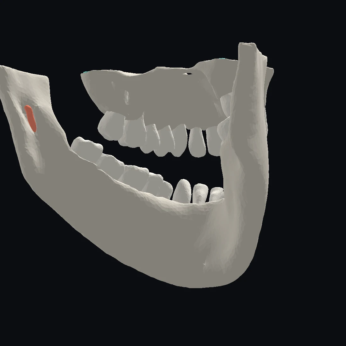
Impacted third molar
Impacted tooth no. 32 with the root apices abutting the right inferior alveolar canal.
Maxillary sinus
The left maxillary sinus demonstrates severe mucosal thickening.
Periodontal bone loss
Generalized moderate periodontal bone loss of the maxillary teeth.
Streamline your workflow. A single shared workspace for dentists and radiologists to manage cases, billing, and communication. Spend less time on administrative overhead and more on patient care.
See how it worksReferral question
Impacted lower left third molar. What is its relation to the inferior alveolar canal?
CBCT · 215 slices uploaded by the imaging partner
Report
Starts when the study is in.
Segmentation, airway analysis, and implant planning sit in the same window as the report, so the study and the write-up never separate.
View demo casesReport
Segmentation and other automated features shown here are in development and have not been cleared by the FDA. Every report is reviewed and signed by a licensed oral and maxillofacial radiologist.
How it works
A referral creates the record. Every question, image, and note stays attached to this single workspace from day one.
Upload your own study or refer a patient for a scan. The images land in the case, ready for immediate review.
Review the volume side-by-side with your report. AI-powered tools assist your measurements, segmentation, and planning.
A CBCT is more than a screenshot.
Sign and return the complete package. The study and the final, signed report go back to the dentist together.
Referral question
Impacted lower left third molar. What is its relation to the inferior alveolar canal?
Awaiting imaging
Report
Starts when the study is in.
Referral question
Impacted lower left third molar. What is its relation to the inferior alveolar canal?
CBCT · 215 slices uploaded by the imaging partner
Report
Starts when the study is in.
Referral question
Impacted lower left third molar. What is its relation to the inferior alveolar canal?
CBCT · 215 slices uploaded by the imaging partner
Report
Referral question
Impacted lower left third molar. What is its relation to the inferior alveolar canal?
CBCT · 215 slices with the signed report attached
Report
For radiologists
Invite your referring dentists. Receive cases, review the imaging, write the report, and return it from one queue.
For radiologistsFor dentists
Add the clinical question and imaging. Track the case and receive the completed report in the same place.
For dentistsNo radiologist yet? Open a free workspace. Free for your practice, with or without a radiologist to send to.
Refer the patient to a Dentica imaging partner. The partner uploads the completed study to the same case, ready for review. Refer a patient for imaging.
Questions
Dentica is the shared workspace where a dental imaging case stays together, from the referral question through the study to the signed report. Reports are reviewed and signed by licensed oral and maxillofacial radiologists.
A licensed oral and maxillofacial radiologist. Dentica provides the workflow software. The clinical work and the signature stay with the radiologist.
No. The case goes to the radiologist you already refer to, under their own name and their own clinical relationship.
Refer the patient to a Dentica imaging partner. The partner uploads the completed study to the same case, so the referral question and the imaging stay together.
Yes. The Sign up button opens the product signup, and the workspace is free to open and use. The software status page in the footer lists what is available today.
About Dentica
Both founders read cone-beam studies for a living, and Dentica is the workflow they already run, built into software a practice can use.